Choose a Skin Cancer Removal Treatment with a Cure Rate of up to 99 Percent for the Little Rock Area, Central Arkansas, and Beyond
Anyone diagnosed with skin cancer will understandably want the most effective treatment available. While modern medicine has given us a number of options, the ideal approach for many patients is a sophisticated excision technique known as Mohs micrographic surgery. The Little Rock area’s Dermatology Group of Arkansas offers this option, which involves the painstakingly methodical removal and examination of tissue in the treatment area. Each step is repeated until all of the cancer cells are gone, providing a cure rate of 95 to 99 percent for cancers that haven’t been previously treated. This is the highest cure rate out of all available options for treating basal cell carcinoma and squamous cell carcinoma. Dermatology Group of Arkansas has two dermatologists on staff who are specially trained and board certified in this advanced micrographic technique,
Mohs surgery is especially good for treating cancerous lesions and tumors that appear small on the surface, but that expand significantly into deeper layers—like an iceberg floating in the ocean. What is visible on top can be just a fraction of the whole. In other words, cancerous cells can often be found beyond what is apparent, exisiting deeper than expected in the skin, as well as developing along vessels, nerves, and cartilage. Less-precise excision procedures can leave cancerous cells behind, which allows the disease to potentially continue to spread. Mohs micrographic surgery, however, is designed to precisely “track” the path of the malignant cells, allowing the surgeon to remove all of the cancerous tissue and maximize the patient’s long-term health.
Contact Us Today
Schedule your consultation for Mohs micrographic surgery in the Little Rock area and beyond. Call us at the Dermatology Group of Arkansas at 501-227-8422.

Is Mohs Micrographic Surgery Best for Everyone?
Researching skin cancer treatments can bring up a lot of information, with options including topical medications, radiation therapy, chemical treatments, and more. Exploring the various treatments is a good idea, as it can help patients to get an idea of what is possible. It is most important, however, to discuss these treatments with a qualified professional who can make the determination of which route would be best for you to take.
Some patients may not need Mohs micrographic surgery for excision, because their cancerous lesion is small, not prone to spreading, and in an area that wouldn’t really benefit from the tissue-sparing technique. Patients who most benefit from the technique tend to be those whose lesion is located on a highly visible or especially sensitive area, such as the nose or near the eyelid.
How Does Mohs Micrographic Surgery Work?
Mohs surgery involves removing a thin layer of tissue, then preparing the sample and examining it under a microscope to look for cancer cells. These steps repeat until no cancerous cells are detected. The procedure is done in stages, including lab work, while the patient waits.
The University of Wisconsin’s Dr. Frederick Mohs first developed Mohs surgery in the 1930s, creating a procedure that involves several specific steps for removing cancer cells without harming the healthy, surrounding tissue.
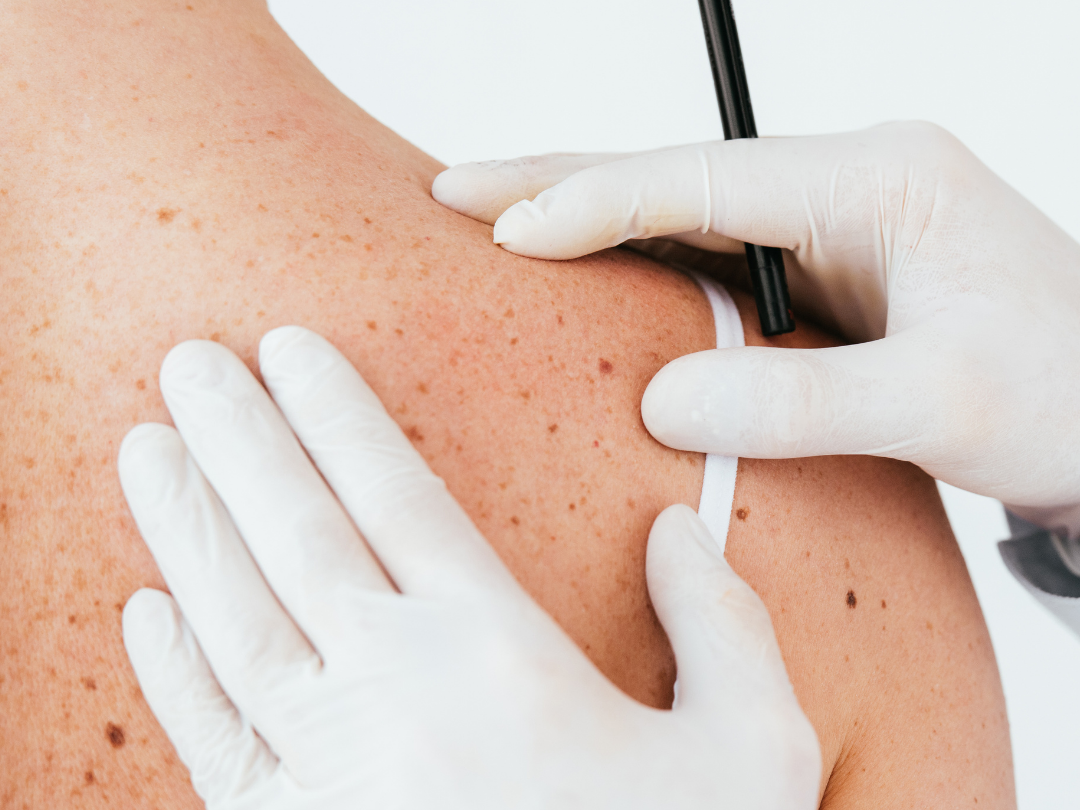
First, the provider indicates the outline of the visible part of the cancer by drawing around it on the skin’s surface.
Next, the treatment area is injected with a local anesthetic to provide a numbing effect.
Once sensation is reduced in the area, the Mohs micrographic surgeon begins the most painstaking aspect of the procedure: removing the cancerous cells in a methodical way. The first step of this phase involves taking the visible cancer and an extra layer of skin from the treatment area. Following this, the patient is bandaged and encouraged to rest comfortably while the surgeon takes the excised layer of skin to the laboratory and processes it into slides by cutting the tissue into sections and staining them.
For the next step, the surgeon carefully checks all of the edges of the skin sample—including the bottom—for telltale signs of cancer cells. The visible results are marked on what is known as a “Mohs map” of the tissue section. If cancer cells are seen, the surgeon will take another sample, but only from the areas where the malignant cells were found.
This process is unique to Mohs micrographic surgery. Equipped with the knowledge of exactly which directions the lesion extends below the surface, the surgeon can follow the map and be sure all traces of malignant cells are being cut out. Since the tissue examinations happen right after the samples are removed, there is no need to wait on anything to come back from an external lab.
The Mohs micrographic surgery process will continue step by step, repeating the back-to-back excision and examination process, until no signs of cancer cells are detected in any of a sample’s margins—including its base. Finding “clear margins” gives the surgeon confidence that all of a cancer’s extensions or “roots” have been excised.
Finally, you will discuss plans to repair the wound made by the Mohs surgery excision. Most of these repairs are handled directly in the Dermatology Group of Arkansas office, where our experienced team performs linear closures, grafts, flaps, and more.
First and foremost, our goal is to entirely remove the cancer to maximize your long-term health. We also know that keeping visible scarring to a minimum is important to our Mohs surgery patients’ self-esteem. Happily, it is quite possible to safely accomplish both.
Because every patient’s cancer develops in a distinct way, there is no fixed length of time to expect a Mohs micrographic surgery to take. The excision and examination process tends to move relatively quickly, but we still encourage patients to set aside a day for the procedure—just in case.
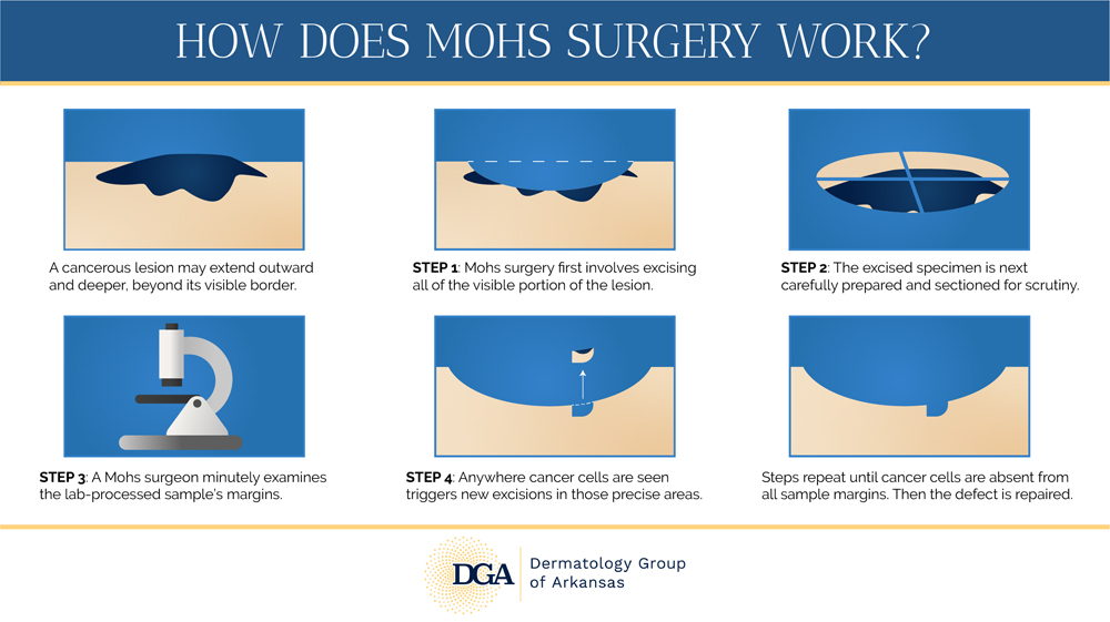
Mohs surgery at the Little Rock area’s Dermatology Group of Arkansas involves several steps, including precise excision of the tumor, preparation of samples, intense scrutiny of samples for signs of remaining cancer cells, and new excisions in necessary areas. Microscopic examinations and excisions continue until there are no signs of skin cancer, after which repair and reconstruction will be discussed.
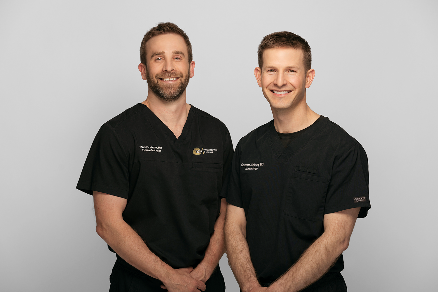
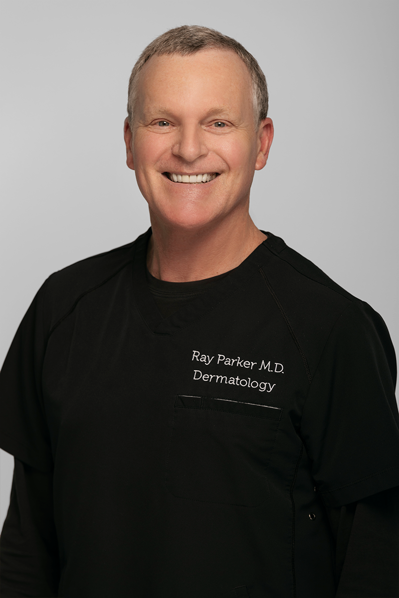
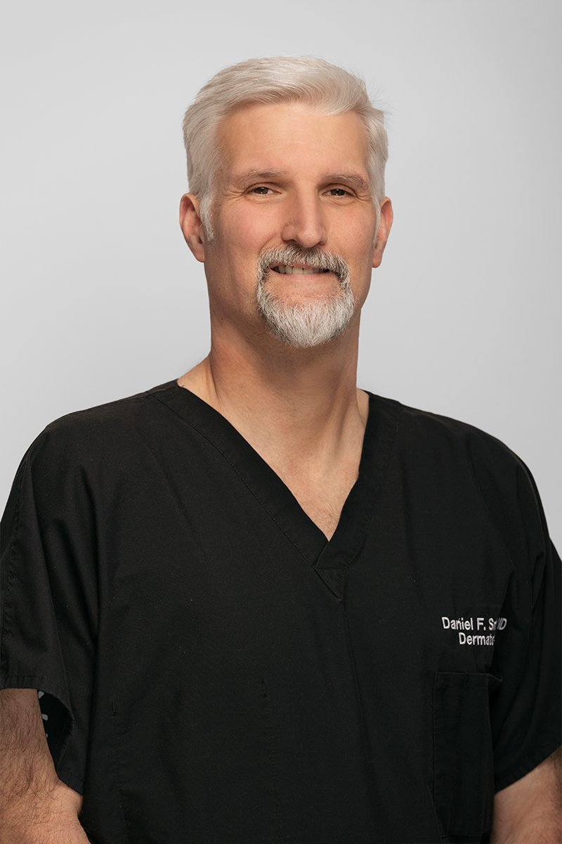
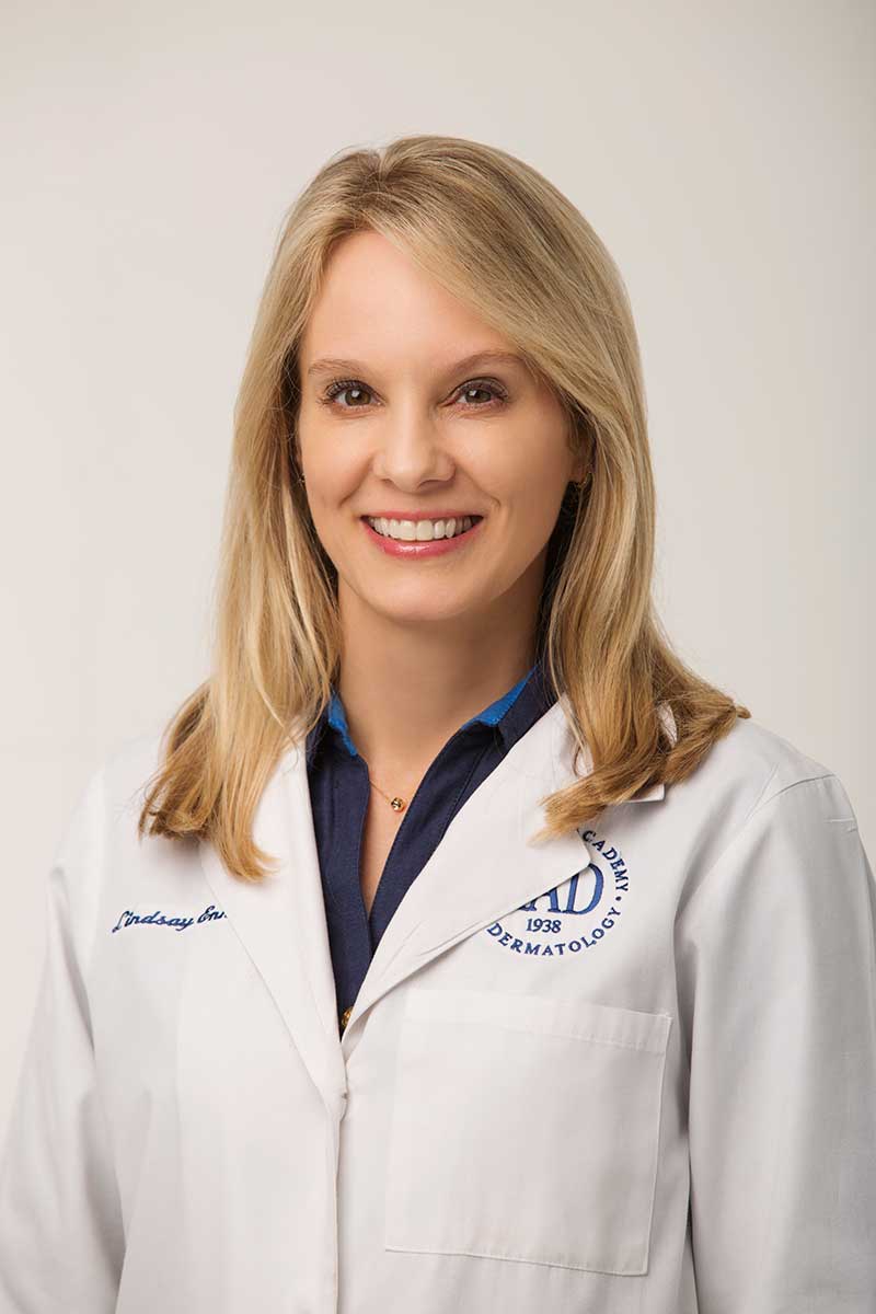
Meet Your Doctors
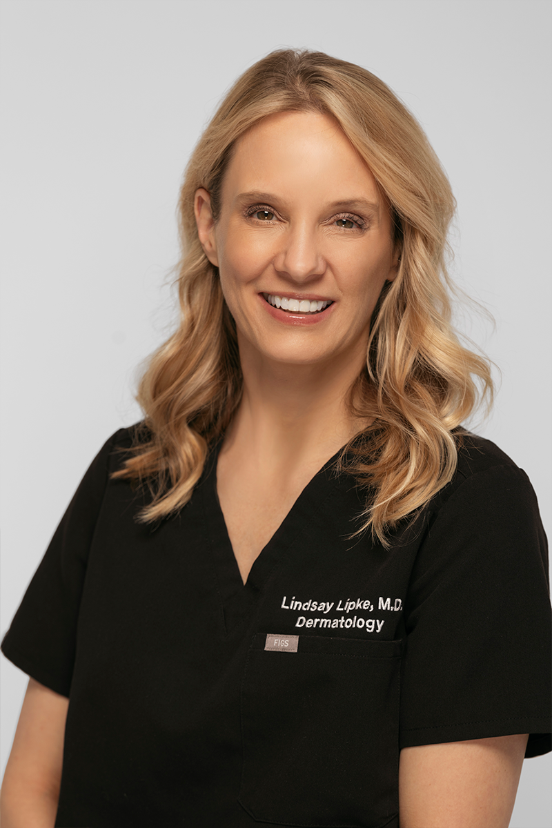
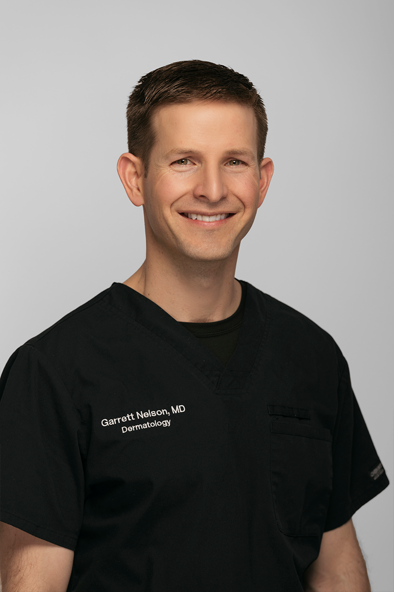
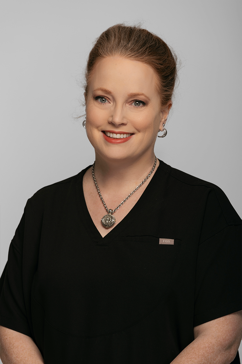
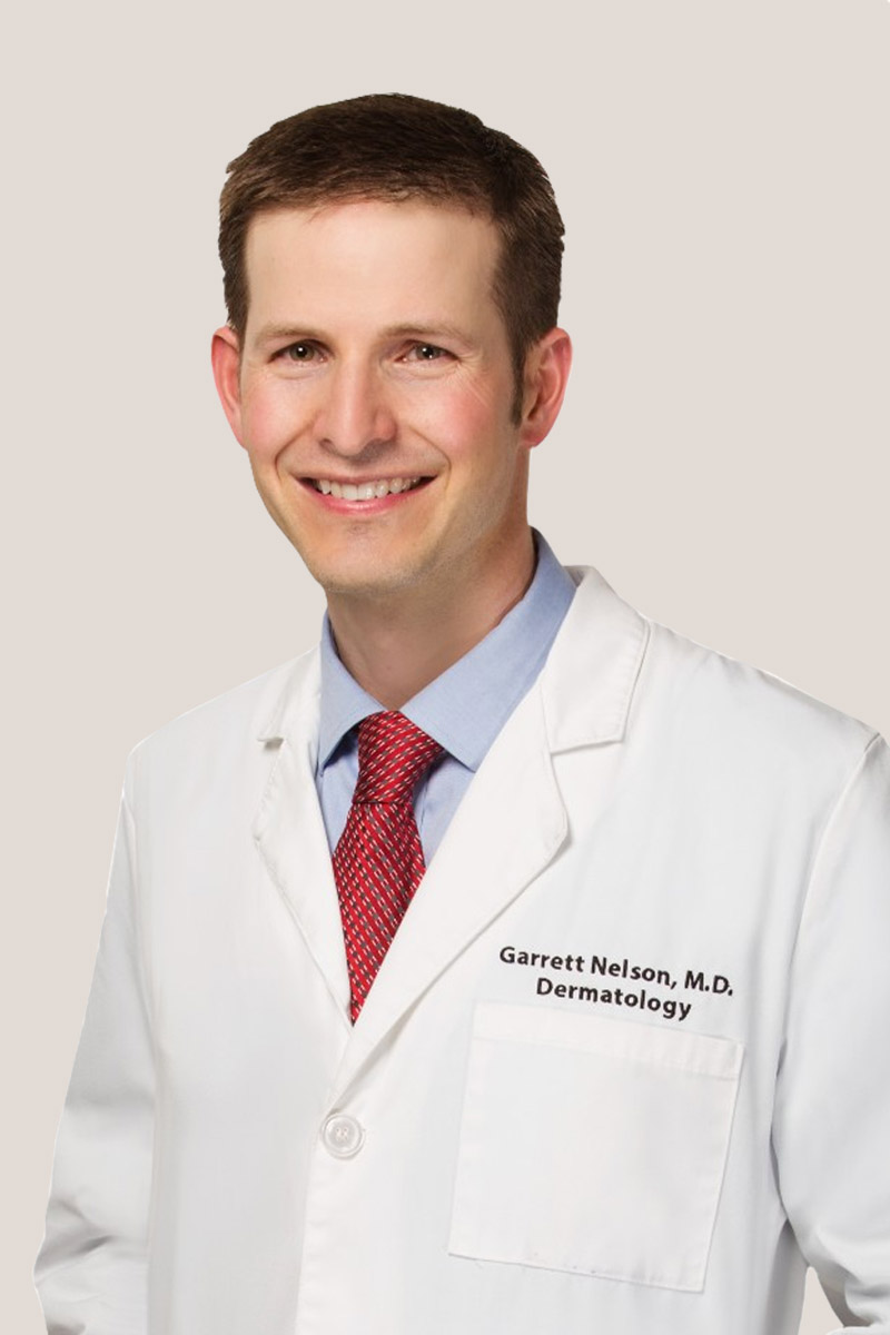
Meet Your Doctors
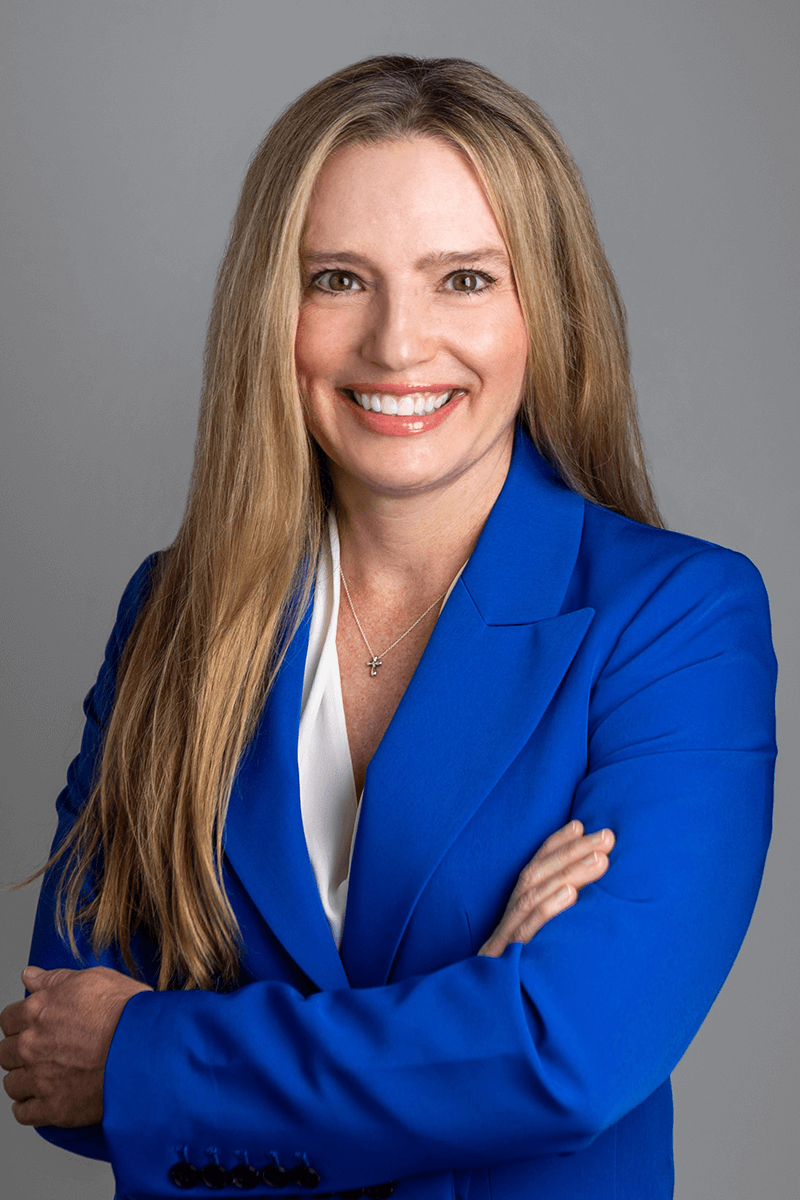
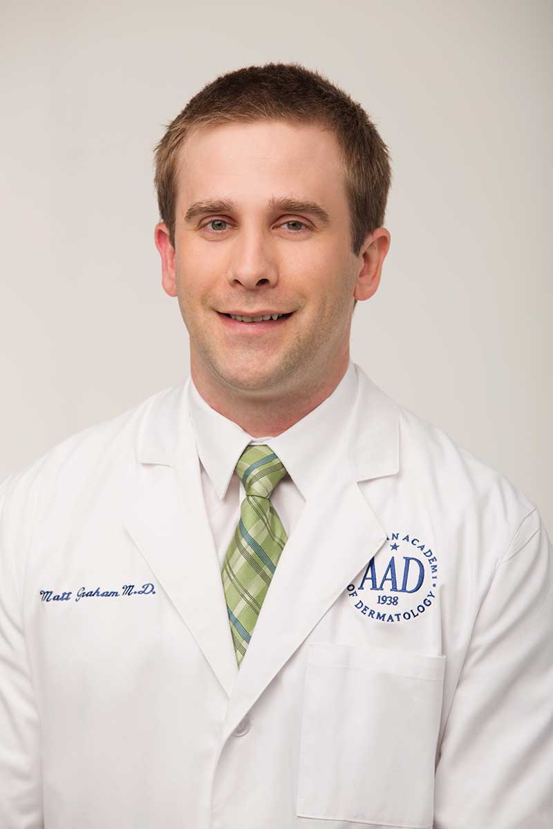
Meet Your Doctors
What Makes Mohs Micrographic Surgery Stand Out from Other Cancer Treatments?
The main appeal of Mohs micrographic surgery is its extremely high cure rate, made possible by the meticulous microscopic checking of all the edges and base of the lesion, which happens throughout the actual procedure.
Another benefit of Mohs micrographic surgery is keeping the surgical wound as small as possible. Only the skin affected by cancerous cells is excised, so a maximum amount of healthy tissue is left intact. This makes damage to important structures near the cancer—such as the eyes—much less of a risk.
Minimizing the wound also means that the scar will be as small as possible once it has healed. Typical excision procedures simply remove the lesion and a chunk of the surrounding tissue, which is not only precise, but also leaves a larger-than-necessary defect.
All of this makes Mohs surgery ideal for treating cancers that develop in skin that is particularly delicate, sensitive, or highly visible. Mohs micrographic surgery patients are often diagnosed with basal or squamous cell cancer found in the skin of highly prominent features: their nose, their ears, or their lips. The surgery also recommended for patients who have cancers on areas that are frequently exposed to the sun, such as the head, back of the neck, and tops of the hands.
There are also numerous other benefits of Mohs micrographic surgery: It is a single-visit, outpatient surgery, so no coming and going is required. Only local anesthesia is required, which does away with the risks and side effects associated with a general anesthetic. It provides precise results for a more accurate treatment and recovery. Healthy tissue is spared, which makes healing and recovery even faster. Finally, the lab work is done on site, so there is no hassle related to the process of sending tissue samples to a different laboratory.
Since the surgeon is able to ensure that all of the skin cancer has been removed, Mohs surgery reduces the need for other types of procedures or additional surgery in the future.
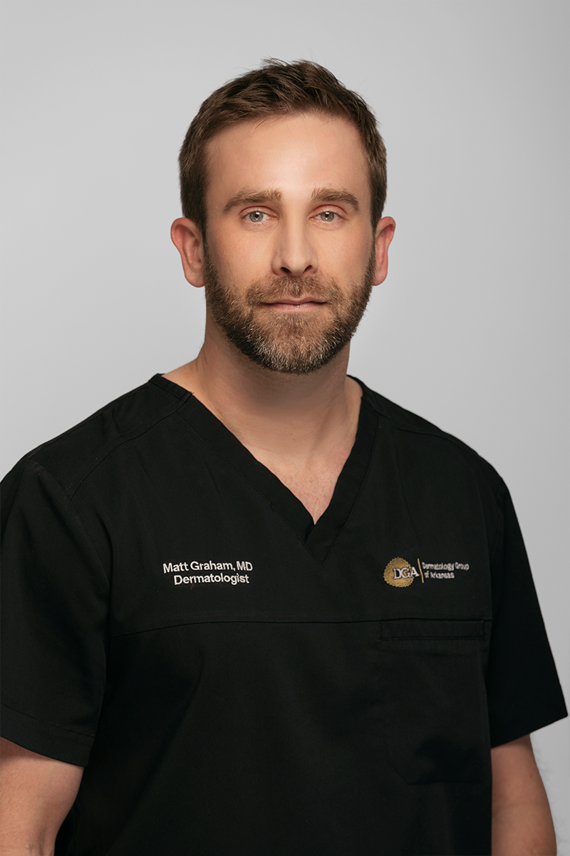
Mohs surgery is a very unique approach to treating certain cancers of the skin, so it requires specialized education and training. At the Dermatology Group of Arkansas, Dr. Garrett Nelson and Dr. Matt Graham are Mohs micrographic surgeons with voluntary board certification and extensive experience in the advanced cancer-removing and tissue-sparing technique.
In addition to our dermatologists specially trained in Mohs micrographic surgery, Dermatology Group of Arkansas also features a highly trained surgical staff, histotechnicians who prepare and study tissue samples, and an on-site lab.
Contact Us Today
Contact the Dermatology Group of Arkansas today if you are interested in learning more about Mohs micrographic surgery in the Little Rock area. Call 501-227-8422.